Talk Overview
This lecture describes several methods for approximately doubling the resolution of the light microscope: 1) illuminating and detecting through two objectives, 2) structured illumination microscopy (SIM) with a patterned light source, and 3) saturating (high intensity) structured illumination to provide further resolution extension. The methods and examples of images are presented.
Questions
- What physical property(ies) limit the resolution of microscopes?
- Wavelength of the detected light
- Numerical aperture of the objective lens
- Magnification of the objective lens
- Wavelength of the light used for fluorescent excitation
- A, B, C, and D
- A and B
- The use of 2 opposing objective lenses can increase resolution by
- Having a larger effective numerical aperture than possible with 1 objective
- Interference of the emitted light collected through the 2 lenses
- Interference of the excitation light going through both lenses
- A, B, C and D
- The resolution of I5m microscopy is best in
- XY (lateral)
- Z (axial)
- Same laterally and axially when using interference
- Structured illumination microscopy:
- Uses patterned light to illuminate the sample
- Produces images that can be directly observed through the eye-piece
- Produces images that can only be observed after computer-reconstruction of the raw images
- Can not be used for fluorescence microscopy
- A and B
- A. and C
- he resolution of Structured Illumination microscopy is best in
- XY (lateral)
- Z (axial)
- Same laterally and axially when using interference
- The resolution of Structured Illumination microscopy
- Is at best twice the resolution of a conventional microscope
- Can be arbitrarily high when using non-linear phenomena
- Depends on the magnification of the objective lens
- Is a function of the computer algorithm used for image reconstruction
- Live cell imaging using structured illumination
- Can only be done using non-linear phenomena such as dye saturation
- Is best executed with a system that can move the illumination pattern very quickly so that the sample does not change in between images
- Does not work
- Results in less photo-bleaching than conventional microscopy
Answers
View AnswersSpeaker Bio
David Agard
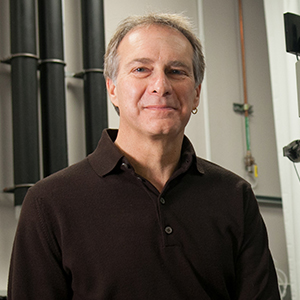
David Agard is Professor of Biochemistry and Biophysics at the University of California, San Francisco, an Investigator of the Howard Hughes Medical Institute and a member of the National Academy of Sciences. Agard’s lab studies the relationship between the structure and the function of macro and supramolecular complexes such as centrosomes, chromosomes, and HSP 90…. Continue Reading


Cheng says
I was wondering if you could share the slides of this talk.