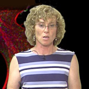
Dr. Waterman is chief of the Laboratory of Cell and Tissue Morphodynamics at the National Heart Lung and Blood Institute, National Institutes of Health and she has been co-director of the Physiology Course at the Marine Biological Laboratory for the past 4 years. Waterman’s lab studies the interactions between actin and focal adhesions, taking advantage of their knowledge of advanced microscopic techniques.

Talks with this Speaker
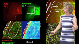
Quantitative Analysis of Speckle Microscopy
Clare Waterman, developer of fluorescent speckle microscopy, describes computational tools (developed by Gaudenz Danuser) for automatic quantitative analysis of speckle microscopy data. (Talk recorded in July 2012)

Audience:
- Researcher
Duration: 6:16
Measuring Dynamics: Fluorescent Speckle Microscopy
Clare Waterman, the inventor of fluorescent speckle microscopy, describes how to prepare samples for speckle microscopy and how to image them. (Talk recorded in July 2012)

Audience:
- Researcher
Duration: 22:50


