Talk Overview
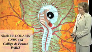
The neural crest (NC) is a transitory structure of the Vertebrate embryo. It forms when the neural tube closes through the epithelio- mesenchymal transition of the cells in the joining neural folds. Its constitutive cells are endowed of migratory capacities and are highly pluripotent. NC cells migrate in the developing embryo along definite pathways, at precise periods of time during embryogenesis and settle in elected sites in the body where they develop into a large of cell types. The paramount role played by the NC in the development of higher Vertebrates has been deciphered in the last decades through the use of cell marking technique that I devised in 1969. This technique is based on the constrution of chimeric embryos between two species of birds the chick (Gallus gallus) and the Japanese quail (Coturnix coturnix japonica) whose cells can be distinguished by the structure of their interphase nucleus or by applying species specific monoclonal antibodies on chimeric tissues.
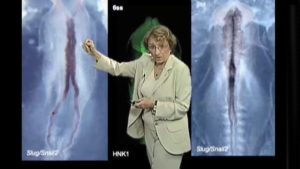
The Q/C (quail/chick) marker system revealed that apart from the peripheral nervous system (PNS), the melanocytes and some endocrine tissues, the NC plays a major role in the construction of the vertebrate head. The entire facial skeleton and part of the skull (frontal, squamosal, parietal bones) are of NC origin together with the connective tissues of the face and ventral part of the neck. The contribution of the NC to the heart and blood vessel wall (except for the endothelial cells) was also demonstrated. This led Gans and Northcutt (1983) to propose that the NC played a critical role in the evolution of Vertebrates since it allowed the formation of a “New Head” and appeared at the transition between Cephalocordates (whose extant representative, the Amphioxius, is devoid of NC) and Vertebrates.
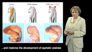
Further studies have shown that the NC cells which participate in facial skeletogenesis correspond to the anteriormost region of the body axis where the genes of the Hox cluster are not expressed. If the forced expression of Hoxa2, Hoxa3 and Hoxb4 (the most anteriorly expressed Hox genes) is induced in this part of the neural fold, brain development is deeply affected with anencephaly and no skeletogenesis takes place in the face which fails to develop. This phenotype is reproduced when the anterior neural fold is surgically removed before NC emigration. One of the early consequence of these experiments concerns the expression of Fgf8 which disappears in the anterior neural ridge (mostly) and in the branchial arch ectoderm. We found that the phenotype resulting from the excision can be rescued by providing the developing head with an exogenous source of Fgf8, thus showing the important role of this signaling molecule in head development. The next study (in progress in my laboratory) concerns the molecular mechanisms through which the NC affects Fgf8 production during cephalogenesis in Vertebrates.
Speaker Bio
Nicole LeDouarin
Nicole Le Douarin is honorary Professor at the College de France in Paris and former Director of the Institut d’Embryologie Cellulaire et Moléculaire du Collège de France et du CNRS. Presently, she works at the Institut Alfred Fessard – CNRS – à Gif-sur-Yvette, France. Nicole Le Douarin understood that a peculiarity of quail cells, that… Continue Reading


Leave a Reply