Talk Overview
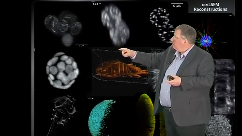
Selective Plane Illumination Microscopy (SPIM) greatly reduces phototoxicity in comparison with other fluorescent imaging modalities and makes it possible to image living small animals in 3D over extended periods of time. This talk describes an extension of SPIM such that it can be used with specimens on a coverslip rather than in a capillary (inverted SPIM or iSPIM), and a second modification that images the specimen using two perpendicular light sheets (Dual-View iSPIM or diSPIM), resulting in 3D datasets with the same resolution in X, Y, and Z (isotropic resolution).
Questions
-
What distinguishes iSPIM from SPIM?
-
What is the advantage of diSPIM over iSPIM?
-
What types of biological objects can be images with diSPIM?
Answers
View AnswersSpeaker Bio
Hari Shroff

Hari Shroff is an Investigator at the National Institute of Biomedical Imaging and Bioengineering at the National Institutes of Health. During his post-doc, which was with Eric Betzig at HHMI’s Janelia Farm Research Campus, he focused on the development of PALM (photoactivated localization microscopy) microscopy. Since then he has developed several microscopy techniques, such as… Continue Reading


Leave a Reply