Talk Overview
Transmission electron microscopy (TEM) offers the possibility of visualizing biological structures at resolution well beyond that of light microscopy. Whether you are interested in the ultrastructure of cells and organelles, or in the detailed molecular structure of biological macromolecules, different modalities of TEM can generally be applied to your system of interest.
The lecture reviews the physical principles underlying image formation by the interaction of electrons with matter, introduces you to basic and advanced instruments and to sample preparation techniques. Using a number of biological examples from work in the Nogales lab, the lecture then describes the capabilities of the TEM methodology. Special emphasis is placed on the image processing methods used to obtain three-dimensional information from TEM data.
Speaker Bio
Eva Nogales
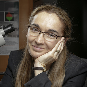
Eva Nogales is a Howard Hughes Medical Institute investigator, a Professor of Molecular Cell Biology at the University of California, Berkeley, and Senior Faculty Scientist at the Lawrence Berkeley National Laboratory. She obtained her B.S. degree in physics from the Universidad Autonoma de Madrid (Spain) and did her thesis work at the Synchrotron Radiation Source… Continue Reading


Bulat says
Thanks a lot Dr. Nogales for a great lecture and a presentation!
I wish there were more videos with further explanation of the theory behind cryo-EM, and the experimental techniques, and tutorials for the software.